Thursday, 20 May 2021

अँजिओप्लास्टी म्हणजे काय ?

        अँजिओप्लास्टी म्हणजे काय ?

दिवसेंदिवस हृदयरोगांचे प्रमाण वाढत आहे. धूम्रपान, व्यायामाचा अभाव, लठ्ठपणा, आहारात चरबी युक्त पदार्थाचे जास्त प्रमाणात सेवन अशा अनेक कारणांमुळे हृदयाला रक्तपुरवठा करणाऱ्या कोरोनरी या रक्तवाहिनीमध्ये अडथळा निर्माण होतो. त्यामुळे हृदयाचा रक्तपुरवठा कमी होऊन श्रमानंतर छातीत दुखणे अशी लक्षणे दिसून येतात. उपचार न घेतल्यास पुढे हार्ट अॅटॅकही येऊ शकतो.

कोरोनरी रक्तवाहिनीतील अडथळा दूर करण्यासाठी व तेथील रक्तपुरवठा सुरळीत करण्यासाठी अँजिओप्लास्टी या तंत्राचा वापर केला जातो. यात मांडीतील वा हातातील रोहिणीतून हृदयरोगतज्ज्ञ कॅथेटर आत टाकतात. या कॅथेटर मधून पाठवण्यात आलेल्या बलूनच्या साहाय्याने रक्तवाहिनीतील अडथळा दूर केला जातो. काही वेळा रक्तवाहिनीची पोकळी तशीच राहावी म्हणून आधार वा स्टेंटही बसवला जातो. याठिकाणी रक्त साकळण्याची क्रिया होऊ नये यासाठी विशिष्ट औषधी द्रव्य सोडणारे स्टेंटही आता उपलब्ध आहेत. आजकाल औषधीयुक्त स्टेटचा वापर जास्त प्रमाणात होताना दिसतो.

अँजिओप्लास्टी ही तशी सोपी उपचार पद्धती आहे. १ ते २ तासात प्रोसिजर पूर्ण होते. रूग्ण या दरम्यान पूर्णत: शुद्धीवर असतो. १ ते २ दिवसात तो घरी जाऊ शकतो. सध्या या उपचाराला १ ते २ लाख रुपये खर्च येत असल्याने ती सर्वसामान्यांच्या आवाक्याबाहेर आहे. अँजिओप्लास्टीनंतर पहिल्या सहा महिन्यात सुमारे ३०% व्यक्तींमध्ये पुन्हा अडवळे निर्माण होऊ शकतात. म्हणून या काळात जास्त काळजी घ्यायला हवी. मुख्य म्हणजे अँजिओप्लास्टीसाठी रुग्णाची निवड अचूकपणे करायला हवी. वैद्यक व्यवसायाचे झालेले बाजारीकरण लक्षात घेता रुग्णांनी असा खर्चिक उपचार घेण्यापूर्वी दोन डॉक्टरांचा सल्ला घेणे श्रेयस्कर ठरेल


Wednesday, 21 April 2021

Human Eye

Human Eye structure and function
                     Human eye  
               Anatomical structure 
       
     The human eye is the organ of sight which is located in deep depression of skull called as orbits. It is attached to the orbit by 6 muscles that allow eyeball to move. Eyeball is spherical and has three layers. The first two 2 layer are mesodermal and the third ectodermal in origin.
1)  Superior rectus muscles  or fibrous tunic/The sclera __ this is the outermost, thick and tough, non vascular covering around the eyeball. Over the projecting parts of eyeball, it is transparent, forming the cornea. It lacks blood vessels but it is composed of special type of connective tissue. It is covered externally by a thin transparent membrane called conjuctiva.
2) lnferior rectus muscle__ distracted muscle fibres are  inserted into sclera for eyeball movements. The 3rd,4th & 6th cranial nerves control the eyeball movements to observe in different directions. Light passes and refracted through  cornea and rays begin to converge.
3) Vitreous gel body __ space between cornea and lens is called aqueous chamber . It is filled with clear watery fluid called aqueous  humour , which is a tissue fluid containing glucose, amino acids, respiratory gases, metabolic wastes and ascorbic acid.
     This vitreous chamber is filled with viscous, jelly like humour containing 99% water, salt, a a little quantity of mucoproteins and hyaluronic acid.lt is the part between lens and retina. 
4)Ciliary body and muscles__  lt is located just behind the junction between the main part if sclera and cornea.lt contains a network of blood vessels supplying food and oxygen to eye. It is deeply pigment and is connected  to a thick structure which have blood vessels and muscle fibres that run in circular direction. Contraction and expansion of ciliary muscles bring about increase and decrease in the size of lens 
5) Iris__lt is called diaphragm of eye because it regulates the size of pupil .lt is thin circular structure made up of connective tissues and muscles that surrounds the pupil. Iris is the forward segment of ciliary body which is pigmented  and opaque.The colour of our eyes is determined by the amount of pigment in the Iris. This part is the visible coloured portion of the eye. Iris are heavily pigmented in people with Brown or black eyes but less pigmented in people with blue eyes.
6) Lens__it is a transparent crystalline structure. It is present anteriorly inside the Iris and is held in position by ligaments of ciliary body.
7) Pupile__the aperture surrounded by the Iris in front of lens is called pupil. The large central aperture of iris is called pupil.The movement of pupil is regulated by muscle fibres of iris..
8) Anterior chamber__ lt is filled with clear watery fluid called aqueous humor .The lacrimal gland is situated in the orbit which   secrets  water ,salt  and bactericidal protein called  lysozyme. The function of tears is to bath the front of eye, washing away in a dust, grit and microorganisms.
9)Cornea__lt is transparent anterior circular  portion present in front of the sclera . Sclera is the outer layer of dense connective tissue.
10)Retina__ It is innermost layer of eye.It is very delicate layer which is sensitive to light. It receives  image. bears light sensitive. lt bears light sensitive photoreceptor cells- rods and cones.
(a)Rods: contain purple coloured photosensitive pigments Rhodopsin which are more responsive to dim light i.e  scotopic vision.
(b)Cones:contain violet coloured photosensitive pigments  which function in bright light i. e. photopic vision.
Cones are of 3 differnt types.
(i) short wavelength: sensitive blue cons.
(ii) medium wavelength: sensitive green cones.
(iii) long wavelength: sensitive red cones.
They are called at photopigments.
11)Fovea__lt is  central pit portion of the retina. It contents only cones but no rods and gives the distinct interpretation of an image.Therefore have greatest visual activity. i.e. resolution . Outside the fovea, processing of visual information can occur within the retina.lateral to fovea ,there is a blind spot which does not contain light sensitive cells.
12)Macula__It is yellowish pigment spot is present lateral to the blind spot.
13)Choroid__it is the middle layer present inside the sclera. which is blue in colour contains network of  blood vessels supplying food and oxygen to eye.
Optic nerve __it is sensory in nature and originate from retina of eye and distribution in optic lobes of brain and function is to give  vision. 
   * Common eye defects*
 
1)Myopia / Near sightedness
      Person has difficulty in seeing distant objects clearly.the person can see near objects clearly. Hence it is also called short sight.Engineer site I the eyeball is too long and the retina is too far  from the lens. The light rays converge at a a. In front of the retina resulting in a  blurred image. Concave lens corrects this condition by bringing the light rays to a focus at a point further back.
2) Hypermetropia/farsightedness
 The person has difficulty in seeing near objects. Hypermetropia is just the opposite of myopia. The eyeball is too short and the retina too cloes to the lens. Light rays strike the retina before they have converged again resulting in a blurred image.
convex lenses saraiki far sighted condition by causing the light rays to converge further forward.
3) Astigmatism
     It is due to irregular cornea or lens.lt causing the image to be out of focus, producing  faulty vision. It is corrected by cylindrical lens. It can be diagnosis by retinoscopy. It is commonly occur in in kids between 5 to 17  years old girls and boys.
4) Presbyopia/Old sight
     It is a defect in accommodation occurring in advancing age. Lens becomes elastic and is not able to focus the image of near object while the distant vision is not impaired. It is also called old age long sightedness. This defect is corrected by using convex lens.
5) Strabismus
   It is commonly known as Squint.the eyeball is somewhat bent on onto a side in its orbit so that the optic axis cannot be directed to the same object. Some extra ocular muscle becomes longer or shorter than the normal. It can be cure by small surgery.
6) Cataract / Safaid Motia 
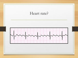
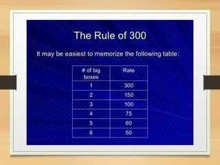
  The lengths loses its transparency and becomes opaque to light and hence vision is impaired.It is corrected by surgical removal of the opaque lens and using biconvex glasses. Intraocular lens implantation is also done. It is commonly occur in old people  &  known as Motibindu.
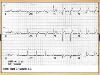
7)Glaucoma/Kala Motia
  It is also more common in older people. If untreated it leads to blindness. The pressure in the anterior cavity of the earth increases to an abnormal level.lt  exerts pressure on the posterior cavity and greatly reduces the blood supply to the retina. Lack of nutrients ultimately damages the Nerve cells of the retina.ek the pressure is due to blockage of the canal of schlemm a new canal can be created surgically.
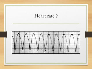
 ** common eye defects such as
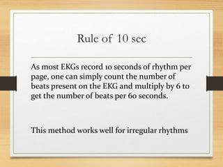
Myopia / Near sightedness ,Hypermetropia/farsightedness,Astigmatism,Presbyopia/Old sight,Strabismus,Cataract / Safaid Motia,Glaucoma/Kala Motia, Conjuctiva, Nightblindness,Colour blindness. दृष्टीदोष_निकटदृष्टिता , दूरदृष्टी ता, विषमदृष्टी, दृष्टीमांद्य , तिरळेपणा, मोतीबिंदू, काच बिंदू, डोळे येणे, रातांधळेपणा, रंगआंधळेपणा.

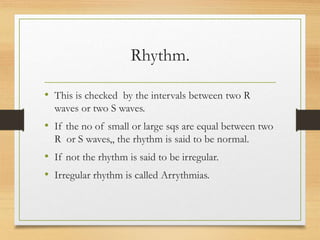
Sunday, 18 April 2021

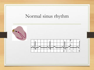
ECG

  1. 1. Electrical Activity of heart
  2. 2. ECG(electrocardiograph) • ECG is define as ‘’ recording of electrical activity of heart on a graph paper.’’ Or • Graphical representation of electrical activity of heart.
  3. 3. Electrocardiograph. • The machine which is used to record the electrical activity of heart is ELECTROCARDIOGRAPH. • a) ECG machine. • b) power lab.
  4. 4. Electrocardiogram. • The graph on which this electrical activity is recorded is called ELECTROCARDIOGRAM.
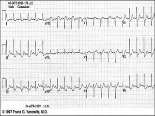
  5. 5. Significance of ECG. • ECG gives information about rate and rhythm of the heart. • The physical orientation of heart i.e axis. • Its a diagnostic tool for various heart conditions like hypertrophies , ischemia, infarction , arrhythmias conduction problems and pace maker activity. • ECG does not provide information about mechanical activity.
  6. 6. ECG paper. • This is long role of paper ,composed of small squares . • One square is 1mm wide and 1 mm high. • On ECG paper there are thick lines, between two thick lines there are 5 small squares. • The speed of ECG machine is 25 mm per second.
  7. 7. ECG paper.
  8. 8. ECG Paper. • The time internal of each small square can be calculated as • 25 small sqrs are equal to 1 second. • 1 small sqr is equal to 1/25 second. • i.e 0.04 seconds.
  9. 9. ECG paper • vertically the small square represent the amount of electrical potential. • One small sqr represent the potential of 0.1 mv. • 10 small squares represent the potential of 1 mv.
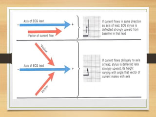
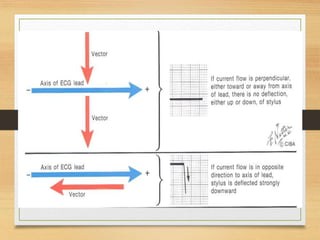
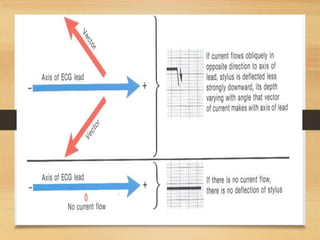
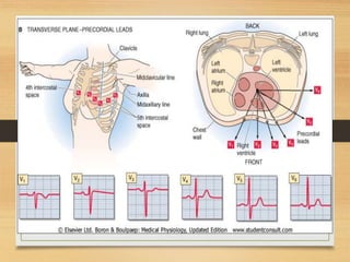
  10. 10. ECG leads. • leads are electrodes which record the electrical potential of heart at different sites. • There are 12 ECG leads. a) 3 bipolar limb leads. b) 3 augmented limb leads.(unipolar). c) 6 chest leads.
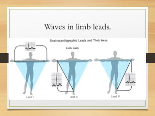
  11. 11. Bipolar limb leads. • Lead 1 = left arm + ve , right arm _ ve • Lead 2 = right arm _ ve ,left leg + ve.. • Lead 3 = left arm _ ve , left leg +ve.
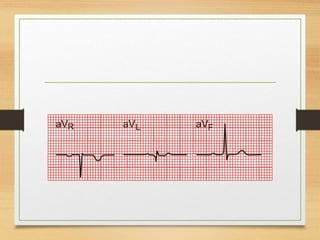
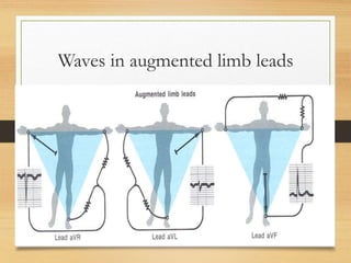
  12. 12. Augmented limb leads. • AVR attach to right arm.. • AVL attach to left arm. • AVF attach to left foot.
  13. 13. Einthoven”s triangle • Einthoven s triangle is a diagrammatic way of illustrating that the two arms and left leg form the apices of triangle surrounding the heart. • It is an equilateral triangle.
  14. 14. Einthoven “s rule. • Einthovens law states that if the electrical potentials of two leads are known at any instant ,the third can be determine by simply summing the first two.
  15. 15. Waveforms in ECG Lecture no 2
  16. 16. Always remember. • Positive electrode is always a reference electrode. • Depolarization wave moves toward the positive electrode gives positive deflection. • Depolarization wave move away from positive electrode gives the negative deflection.
  17. 17. Wave forms.  P wave  Atrial depolarization  QRS complex  Ventricular depolarization  T wave  Ventricular repolarization
  18. 18. Basic terminologies  Base line : flat, straight and isoelectric line  Wave form :deviation or movement away from base line may be upward or downward  Segment : A line between two waves  Interval : a wave form plus a segment this shows time duration  Complex : combination of several wave form without segment.
  19. 19. P wave • P Wave shows atrial depolarization.. • Its duration is 0.1 sec (2 and half small sqr) and height is 2.5 mv (2 and half small sqr). • Presence of p waves in ECG strip shows the sinus rhythm.
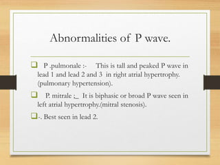
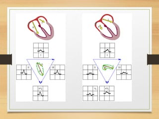
  20. 20. Abnormalities of P wave.  P .pulmonale :- This is tall and peaked P wave in lead 1 and lead 2 and 3 in right atrial hypertrophy. (pulmonary hypertension).  P. mitrale ;_ It is biphasic or broad P wave seen in left atrial hypertrophy.(mitral stenosis). -. Best seen in lead 2.
  21. 21. P wave. • Inverted P wave :- may be present in . • a) Normally in lead V 1 and V2.. • b) In dextro cardia. • c) Incorrectly placed electrodes.
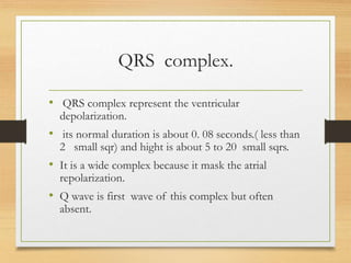
  22. 22. QRS complex. • QRS complex represent the ventricular depolarization. • its normal duration is about 0. 08 seconds.( less than 2 small sqr) and hight is about 5 to 20 small sqrs. • It is a wide complex because it mask the atrial repolarization. • Q wave is first wave of this complex but often absent.
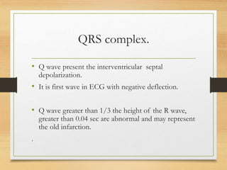
  23. 23. QRS complex. • Q wave present the interventricular septal depolarization. • It is first wave in ECG with negative deflection. • Q wave greater than 1/3 the height of the R wave, greater than 0.04 sec are abnormal and may represent the old infarction. .
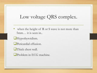
  24. 24. Low voltage QRS complex. • when the height of R or S wave is not more than 5mm… it is seen in.. Hypothyroidism. Pericardial effusion. Thick chest wall. Problem in ECG machine.
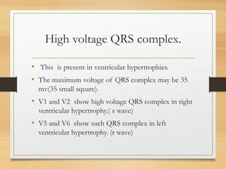
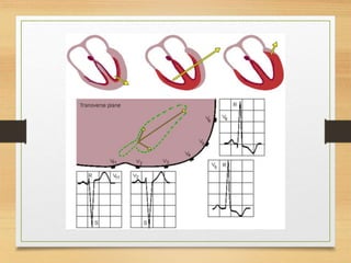
  25. 25. High voltage QRS complex. • This is present in ventricular hypertrophies. • The maximum voltage of QRS complex may be 35 mv(35 small square). • V1 and V2 show high voltage QRS complex in right ventricular hypertrophy.( s wave) • V5 and V6 show such QRS complex in left ventricular hypertrophy. (r wave)
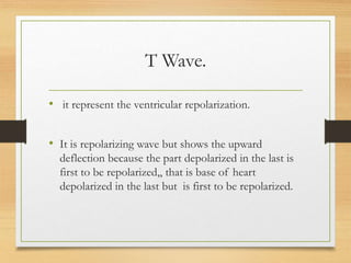
  26. 26. T Wave. • it represent the ventricular repolarization. • It is repolarizing wave but shows the upward deflection because the part depolarized in the last is first to be repolarized,, that is base of heart depolarized in the last but is first to be repolarized.
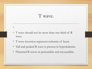
  27. 27. T wave. • • T wave should not be more than one third of R wave. • T wave inversion represent ischemia of heart. • Tall and peaked R wave is present in hyperkalemia. • Flattened R waves in pericarditis and myocarditis.
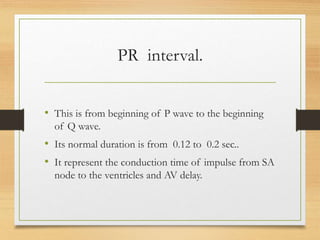
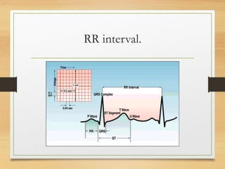
  28. 28. PR interval. • This is from beginning of P wave to the beginning of Q wave. • Its normal duration is from 0.12 to 0.2 sec.. • It represent the conduction time of impulse from SA node to the ventricles and AV delay.
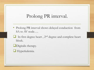
  29. 29. Prolong PR interval. • Prolong PR interval shows delayed conduction from SA to AV node….  In first degree heart , 2nd degree and complete heart block. Digitalis therapy.  Hyperkalemia.
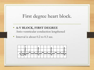
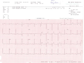
  30. 30. First degree heart block. • A-V BLOCK, FIRST DEGREE Atrio-ventricular conduction lengthened • Interval is about 0.2 to 0.3 sec.
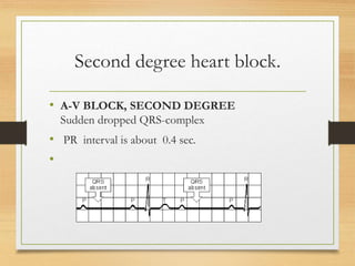
  31. 31. Second degree heart block. • A-V BLOCK, SECOND DEGREE Sudden dropped QRS-complex • PR interval is about 0.4 sec. •
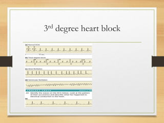
  32. 32. 3rd degree heart block
  33. 33. 3rd degree heart block. • When there is AV block , atria continue to beat at normal rhythem while new pace maker develops in purkinjie system with a rate of 15 to 50 beats /min. • With a sudden block purkinje system cannot take over pace maker activity immediately ,it takes about 16 to 30 sec. during which ventricles fail to contract and person faint. • This delayed pickup of heart beat is called stokes Adams syndrome.
  34. 34. QT interval • Measured from beginning of Q to the end of the T wave . Its duration is about (10 small sqrs). . it indicates total systolic time of ventricles.
  35. 35. RR interval.
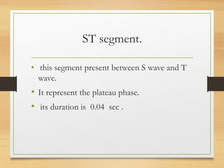
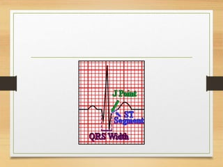
  36. 36. ST segment. • this segment present between S wave and T wave. • It represent the plateau phase. • its duration is 0.04 sec .
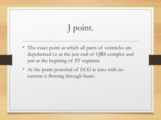
  37. 37. J point. • The exact point at which all parts of ventricles are depolarized i.e at the just end of QRS comples and just at the begining of ST segment. • At the point potential of ECG is zero with no current is flowing through heart.
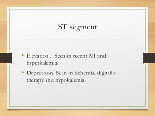
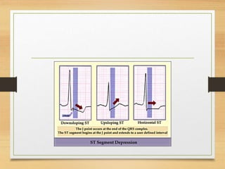
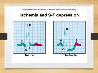
  38. 38. ST segment • Elevation . Seen in recent MI and hyperkalemia. • Depression. Seen in ischemia, digitalis therapy and hypokalemia.
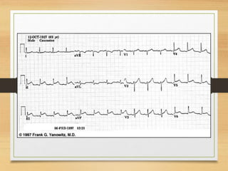
  39. 39. ECG expaination
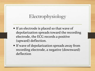
  40. 40. Electrophysiology  If an electrode is placed so that wave of depolarization spreads toward the recording electrode, the ECG records a positive (upward) deflection.  If wave of depolarization spreads away from recording electrode, a negative (downward) deflection
  41. 41. Waves in limb leads.
  42. 42. Waves in augmented limb leads
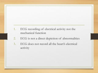
  43. 43. 1. ECG recording of electrical activity not the mechanical function 2. ECG is not a direct depiction of abnormalities 3. ECG does not record all the heart’s electrical activity
  44. 44. ECG
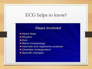
  45. 45. ECG helps to know?
  46. 46. How to calculate heart rate. heart rate define as the no of heart beats per minute.it can be determine by ECG VIA TWO RULES.  Rule of 300  10 Second Rule
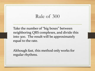
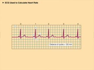
  47. 47. Rule of 300 Take the number of “big boxes” between neighboring QRS complexes, and divide this into 300. The result will be approximately equal to the rate. Although fast, this method only works for regular rhythms.
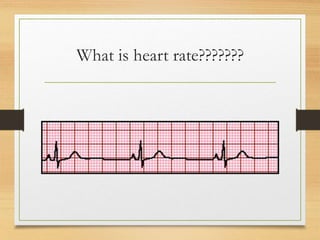
  48. 48. What is heart rate???????
  49. 49. Heart rate?
  50. 50. Heart rate ?
  51. 51. Rule of 10 sec As most EKGs record 10 seconds of rhythm per page, one can simply count the number of beats present on the EKG and multiply by 6 to get the number of beats per 60 seconds. This method works well for irregular rhythms
  52. 52. Rhythm. • This is checked by the intervals between two R waves or two S waves. • If the no of small or large sqs are equal between two R or S waves,, the rhythm is said to be normal. • If not the rhythm is said to be irregular. • Irregular rhythm is called Arrythmias.
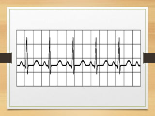
  53. 53. Normal sinus rhythm
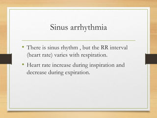
  54. 54. Sinus arrhythmia • There is sinus rhythm , but the RR interval (heart rate) varies with respiration. • Heart rate increase during inspiration and decrease during expiration.
  55. 55. Cardiac Rhythm: Supraventricular
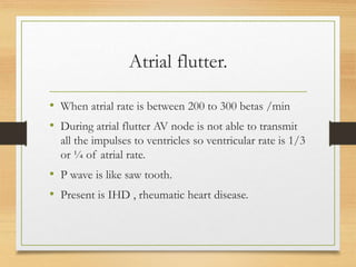
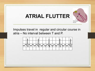
  56. 56. Atrial flutter. • When atrial rate is between 200 to 300 betas /min • During atrial flutter AV node is not able to transmit all the impulses to ventricles so ventricular rate is 1/3 or ¼ of atrial rate. • P wave is like saw tooth. • Present is IHD , rheumatic heart disease.
  57. 57. ATRIAL FLUTTER Impulses travel in regular and circular course in atria – No interval between T and P.
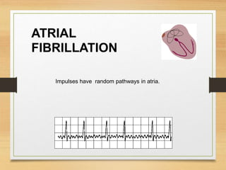
  58. 58. Atrial fibrillation. • A totally irregular and rapid atrial rate in which there is a contraction of only small portion of atrial musculature. • P waves replace by irregular F waves , bcoz whole atria is not contracting. • QRS complex and T waves r normal but rhythm is irregular.
  59. 59. ATRIAL FIBRILLATION Impulses have random pathways in atria.
  60. 60. VENTRICULAR TACHYCARDIA • Impulse originate at ventricular pacemaker – odd/wide QRS complex - often due to myocardial infarction , rate is about 120/ to 220/ min.
  61. 61. Ventricular fibrillation. • Rapid ,irregular and ineffective contractions of small segments of ventricles is termed as ventricular fibrillation. • Peripheral pulse is absent bcoz cardiac out put is zero.
  62. 62. VENTRICULAR FIBRILLATION hapazerd ventricular depolarization – ineffective at pumping – death within minutes • •
  63. 63. Cardiac axis • The flow of current from one point to another is representing by an arrow. This is called Vector. • The direction of flow of current i.e the arrow point is always towards the positive direction. The range of cardiac axis is 0 to +90 degrees. Beyond this limit it is said to be deviated.
  64. 64. Vector Analysis
  65. 65. QRS axis. By near-consensus, the normal QRS axis is defined as ranging from -30° to +90°. -30° to -90° is referred to as a left axis deviation (LAD) +90° to +180° is referred to as a right axis deviation ji(RAD)
  66. 66. Axis deviation • Deviation to R: increased activity in R vent. – obstruction in lung, pulmonary emboli, some heart disease • Deviation to L: increased activity in L vent. – hypertension, aortic stenosis, ischemic heart disease
  67. 67. The quadrant approach
  68. 68. RAD Negative in I, positive in AVF

Project Report on Viral Diseases

Project Report on Viral Diseases Introduction towards Project Report on Viral Diseases : A virus, in the words of one eminent scientist, can...